Bone Cross Section Microscope - Tissue Biomechanics Bme 615 - Accuracy of the tested digitization method was expressed by.
byAnne Hawkins•
0
Bone Cross Section Microscope - Tissue Biomechanics Bme 615 - Accuracy of the tested digitization method was expressed by.. Hope you enjoy and please. File:earthworm crosssection stained microscope slide labeled.jpg. A cross section of a human long bone. Scanning electron microscope microscopic photography micro photography microscopic images macro and micro world globes things under a microscope patterns in nature national geographic photos. Thus as usual microscopic cross sections are experimentally measured.
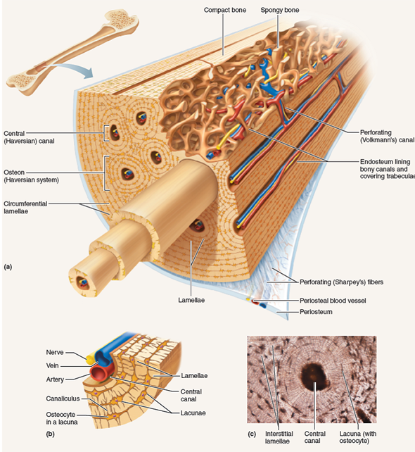
Bone marrow aspiration uses a hollow needle to remove a small sample (about 1 ml) of bone marrow for examination under a microscope. A cross section of a compact bone shows concentric circles called lamellae. Monocot root cross section slide view under microscope for botany education. They build the entire picture, improve your understanding, consolidate the information and facilitate recall. These bone cells have long branching arms (d) which lets them communicate with.
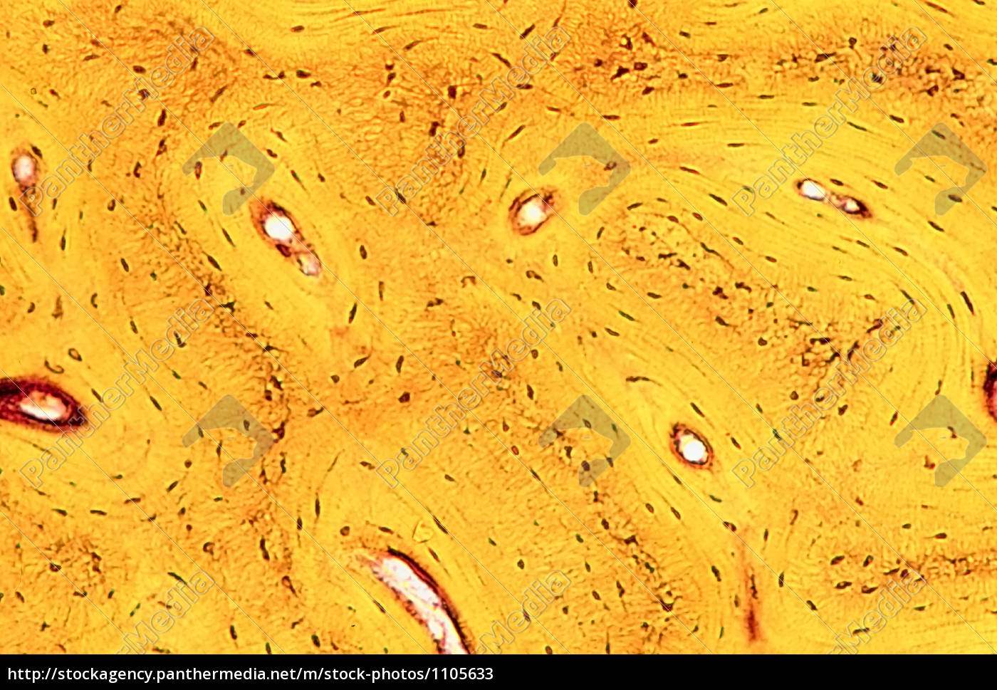
Bone Tissue Of Humans Stock Photo 1105633 Panthermedia Stock Agency from mh-2-stockagency.panthermedia.net Both types of bone marrow are enriched with blood vessels and capillaries.2. They build the entire picture, improve your understanding, consolidate the information and facilitate recall. The microscopic cross section measures the probability of occurrence of a particular nuclear reaction. We obtained 24 axial slices of the normal brain. Bone marrow aspiration uses a hollow needle to remove a small sample (about 1 ml) of bone marrow for examination under a microscope. Jump to navigation jump to search. Hope you enjoy and please. Use electromagnets to focus electrons resulting in significantly greater magnifications and resolutions.
Single, prepared microscope slide of cross section and longitudinal section of a bone.
Cross section performed on focused electon beam (fib) microscope at the university of kentucky's electron microscopy center. When the light that enters the condenser is polarized by placing a polarizer in the filter holder and a second, crossed polarizer at the image plane. The large dark spots are passages for blood vessels and nerves. The microscopic bone cross section image acquired by using electronic microscope and is shown in fig.2. Jump to navigation jump to search. The finished bone section will be bonded to a microscope slide and so the first step is to grind flat and polish the part of the bone that will be glued to the slide. From wikimedia commons, the free media repository. Both types of bone marrow are enriched with blood vessels and capillaries.2. Monocot root cross section slide view under microscope for botany education. In the last decade, considerable technological improvements have been made to repair damaged bones and tissue, such as bone cross sections with implants for microscopic examinations. A cross section of a compact bone shows concentric circles called lamellae. Structural parts of a microscope and their functions. Stained for better visualization of characteristic structures.
The microscopic cross section measures the probability of occurrence of a particular nuclear reaction. This simply involves placing a section of the bone on the microscope stage and viewing. Use electromagnets to focus electrons resulting in significantly greater magnifications and resolutions. The finished bone section will be bonded to a microscope slide and so the first step is to grind flat and polish the part of the bone that will be glued to the slide. Microscope cross section (page 1).
Examining The Microscopic Structure Of Compact Boneif A Mo Chegg Com from media.cheggcdn.com Thin section of dinosaur bone. Structural parts of a microscope and their functions. Compact bone cross section courtesy: We obtained 24 axial slices of the normal brain. A cross section of a compact bone shows concentric circles called lamellae. Jump to navigation jump to search. Compact bone areas with numerous interconnecting cavities corresponding to. Bone marrow aspiration uses a hollow needle to remove a small sample (about 1 ml) of bone marrow for examination under a microscope.
Microscope cross section (page 1).
They build the entire picture, improve your understanding, consolidate the information and facilitate recall. When the light that enters the condenser is polarized by placing a polarizer in the filter holder and a second, crossed polarizer at the image plane. Important features in the bone cross section such as harvesian canals, osteons, osteon fragments, lamellar bone, bony trabeculae, myxoid matrix and artifact for. Hope you enjoy and please. We obtained 24 axial slices of the normal brain. From wikimedia commons, the free media repository. Use electromagnets to focus electrons resulting in significantly greater magnifications and resolutions. Jump to navigation jump to search. Compact bone cross section courtesy: A cross section of a compact bone shows concentric circles called lamellae. The concept of a nuclear cross section can be quantified physically in terms of characteristic area where a larger area means a larger probability of interaction. Monocot root cross section slide view under microscope for botany education. An mri was performed on a healthy subject, with several acquisitions with different weightings:
Thus as usual microscopic cross sections are experimentally measured. Single, prepared microscope slide of cross section and longitudinal section of a bone. This simply involves placing a section of the bone on the microscope stage and viewing. We obtained 24 axial slices of the normal brain. Stained for better visualization of characteristic structures.
Bone Wikipedia from upload.wikimedia.org Compact bone areas with numerous interconnecting cavities corresponding to. Sometimes referred to as 'spongy bone' or 'trabecular bone', cancellous bone is found within the middle of large bones. Accuracy of the tested digitization method was expressed by. An mri was performed on a healthy subject, with several acquisitions with different weightings: This simply involves placing a section of the bone on the microscope stage and viewing. This is a short tutorial using blender 2.8 that shows how to create a bone cross section and using images to create the textures. Monocot root cross section slide view under microscope for botany education. Important features in the bone cross section such as harvesian canals, osteons, osteon fragments, lamellar bone, bony trabeculae, myxoid matrix and artifact for.
Jump to navigation jump to search.
Jump to navigation jump to search. An mri was performed on a healthy subject, with several acquisitions with different weightings: They build the entire picture, improve your understanding, consolidate the information and facilitate recall. Cross section performed on focused electon beam (fib) microscope at the university of kentucky's electron microscopy center. Both types of bone marrow are enriched with blood vessels and capillaries.2. The concept of a nuclear cross section can be quantified physically in terms of characteristic area where a larger area means a larger probability of interaction. The microscopic cross section measures the probability of occurrence of a particular nuclear reaction. Accuracy of the tested digitization method was expressed by. Structural parts of a microscope and their functions. The finished bone section will be bonded to a microscope slide and so the first step is to grind flat and polish the part of the bone that will be glued to the slide. Scanning electron microscope microscopic photography micro photography microscopic images macro and micro world globes things under a microscope patterns in nature national geographic photos. We obtained 24 axial slices of the normal brain. A cross section of a compact bone shows concentric circles called lamellae.
When the light that enters the condenser is polarized by placing a polarizer in the filter holder and a second, crossed polarizer at the image plane bone cross section. In the last decade, considerable technological improvements have been made to repair damaged bones and tissue, such as bone cross sections with implants for microscopic examinations.